Gene Anatomy
 Drs. Huda Akil and Stanley Watson at UM study the relationship between genes, behavior and the brain. Drs. Akil and Watson aim to elucidate and validate the function(s) of genes that demonstrate significant differences in expression or structure and to determine their distribution in pathways, circuits and target systems. Several tools are used in these studies. mRNA in situ hybridization (ISH) histochemistry is performed at UM and UCD to determine the anatomical and cellular distribution of discovered genes. Selected genes are further characterized by determining the structure and function of translated proteins and their regional and cellular distribution. Finally, laboratory experiments using genetically modified animals and cell cultures are conducted at UM, UCD and Stanford to elucidate the role of certain genes in specific brain circuits involved in psychotic and affective processes.
Laser Capture Microscopy
The University of Michigan has two Arcturus Molecular Devices Autopix Laser Capture Microdissection machines, one equipped with epifluorescence filters. The AutoPix LCM system captures brain regions or individual cells onto CapSure Macro LCM caps with laser settings in the mW and msec range.
Confocal Microscopy
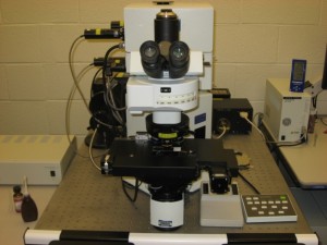
The University of Michigan has two Olympus Flouview 1000 spectral laser-scanning confocal microscopes, one inverted and one upright. The microscopes are built on an Olympus BX61 computer driven microscope and equipped with a Ludl motorized X-Y stage connected to a PC running Olympus FluoView Ver1.5 software. Laser scans of 0.5μm serial Z-section planes can be visualized using a 63x objective.
Microscopy Core
For fluorescence imaging and section analysis, the Molecular and Behavioral Neuroscience Laboratories at Cornell Univeristy use a Nikon E600 microscope with a Microfire digital camera (Optronics), fitted with a computer with 3D motorized stage (Ludl), and Stereo Investigator software (Microbrightfield).
The University of Michigan has a Leica DMR stereology microscope equipped with an Orca Flash 4.0 LT monochrome camera (low-noise CMOS chip, 2048×2048 resolution) and a Lumenera Lt665R color camera (high resolution CCD chip, 2752×2192 resolution). There is also a Zeiss Axiophot stereology microscope equipped with an Optronics MicroFire color camera. Both systems have an XYZ motorized stage, fluorescence imaging capabilities, and use the latest versions of MBF Stereo Investigator and Neurolucida.
For imaging of large tissue volumes (up to 1 cm x 1 cm) that have been optically cleared by CLARITY or other clearing methods, MBNI has a custom built lightsheet microscope (COLM, or Clarity Optimized Lightsheet Microscope). This system is capable of imaging an entire mouse brain in 3-4 hours at half micron resolution. Components include an Omicron SOLE-6 laser engine (405, 488, 515, 561, 594, 647 nm), Hamamatsu Orca Flash 4.0 V2 camera, Olympus 10x (0.60 NA) and 25x (1.00 NA) long working distance CLARITY detection objectives. Custom sample chamber allows for sample and objective to be immersed in refractive index matching solution, maximum 16 mm working distance. FPGA logic is used to control and synchronize various components of the microscope, allowing for high imaging speed and fast data acquisition/transfer. COLM system is controlled by custom-built PC with 196 GB RAM, Intel Xeon E5 2.4 Ghz CPU, Quadro K4200 GPU, 8 TB local SSD storage.
Equipment for Immunohistochemistry
For tissue sectioning, the Molecular and Behavioral Neuroscience Laboratories at Cornell Univeristy use a Leica SM2000R microtome and a Leica VT100 vibratome.

